")
")


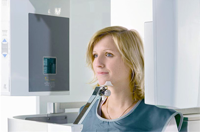
Diagnostics and 3d x-ray technology
We use digital X-rays in our practice. This gives us unique advantages in diagnostics and digital treatment planning.
Using 3D X-ray technology, we are able to look at the roots, precisely evaluate dental and skeletal alignment, and create optimal treatment plans.
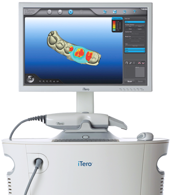
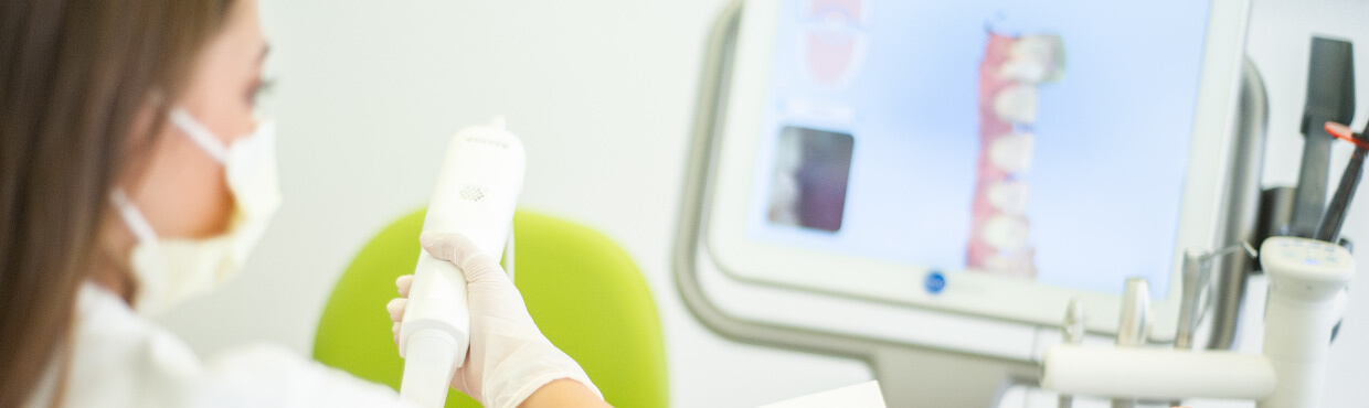
Digital Scanning
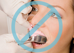
Our practice uses the iTero digital scanner for treatment planning. The advantage of digital scanning is that you don’t have to go through the unpleasant process of having impressions made.
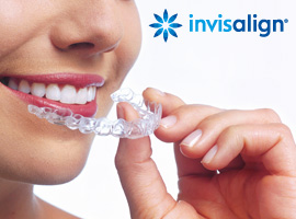
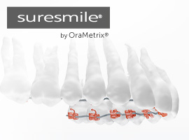
We use the scanner for Invisalign and suresmile , among other treatments..
Advantages
- No conventional impressions
- No gagging
- Precise digital treatment planning
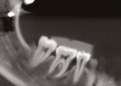
Digital X-Rays
Understanding the exact position of teeth and anatomical structures
Precise information allows for less invasive surgery, for example when a tooth needs to be extracted, and enables better strategies for orthodontic treatment when teeth need to be moved.
Temporomandibular Joint Diagnostics with Digital Volume Tomography
Using 3D X-Ray Technology in TMJ Diagnostics
Using digital volume tomography, we can display 3D X-ray images of the condyles (joint surfaces) and surrounding structures. This allows us to completely analyse the skeletal morphology of the temporomandibular joint, the joint cavity, and the joint functionality, as well as parameters of craniomandibular dysfunction which are critical for treatment and aftercare. With a high-speed scan, the joint can be quickly and precisely imaged even with an open mouth.
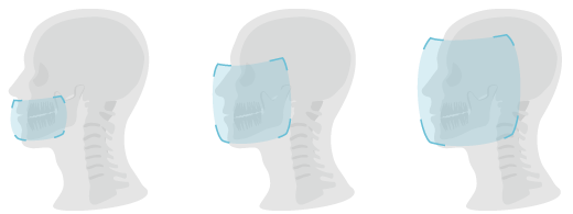
8 cm ø x 8 cm
16 cm ø x 13 cm
23 cm ø x 17 cm
Image Volume
| Mode | Image- Volume | Scan Speed | Voxel Size |
|---|---|---|---|
| Standard | 16 cm Ø x 13 cm | 8,5 s | 0,4 or 0,3 mm |
| High Resolution | 16 cm Ø x 13 cm | 26 s | 0,2 mm |
| Ultra-high Resolution | 8 cm Ø x 8 cm | 26 s | 0,125 mm |
| Cephalometric Mode | 23 cm Ø x 17 cm | 8,5 s | 0,4 mm |
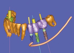
Applications of 3D X-Ray Technology in Orthodontics
- A panorama view from the inside
- Cross sections in all three planes down to 0.125 mm slice thickness
- 3D depiction of all osseous structures
- 3D depiction of all soft tissue structures
- Precise 3D diagnostics of malaligned teeth
- Relative position of the nerve for malaligned teeth in the lower jaw
- 3D examination of the existing bone mass for gap closure
- Buccal and lingual assessment of the bony support of the roots as recession prophylaxis
- Determining the shape of the dental arch depending on the existing 3D bone structure
- 3D planning of orthognathic surgery
- Skeletal anchorage pin planning
- Evaluating the sinuses
- Evaluating the nasal septum
- 2D and 3D cephalometric analysis
State-of-the-art X-ray technology and 3D volume tomography
for dentists, orthodontists, MCG doctors, and ENT specialists.
We accept referrals from colleagues to aid in the diagnosis of their patients
- Comprehensive diagnostic tools
- Identifying malaligned teeth, such as wisdom teeth
- Implant planning
- Evaluating root pockets and bone pockets for endodontics and periodontics
- Surgery planning
Make use of our service and experience
In our practice, we use the KaVo Pan eXam Plus
Send your patients to us for diagnostics and take advantage of our 3D X-ray imaging for your treatment planning.
If you have any questions, don’t hesitate.
We are happy to help.